

6. Co-administration of AHCC and GCP
Studies have demonstrated that GCP has its main functions in anti-angiogenesis and inducing apoptosis to tumor cells and tissues. On another hand, AHCC can increase the immunoreactivity to up-regulate several cytokines such as IL-1, IL-2, IL-l2, IFN-y, TNF - and to enhance the activities of macrophage, LAK and NK cells. Because both substances of AHCC and GCP showed anti-tumor activities, we investigated the synergistic anti-tumor effects of the co-administration of GCP and AHCC in vitro and in vivo. Also, prostate cancer is one of the most difficult cancers in men to be controlled, we used one of human prostate cancer cell lines-PC-3 as the target cell in the study.
1) The growth curves of PC-3 treated with AHCC, GCP or AHCC plus GCP
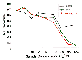
Fig. 7 The growth curves of PC-3 treated with AHCC, GCP or
AHCC plus GCPPC-3 cell line was cultured in 96 well plate in RPMI1640 medium containing 10% FBS at the cell number of 20,000 per ml medium for 24 hrs, then samples dissolved in DMSO (10%) were added to the medium to the final concentrations as shown in the figure. The control was added the same volume of DMSO. After the cells were cultured for another 48 hrs, the growth of the cells was measured using MTT assay kit and the values shown in the figure are means of triple wells.
As seen from Fig. 7, the co-administration of AHCC plus GCP showed a clear dose-dependent inhibitory effect on PC-3 tumor cells.
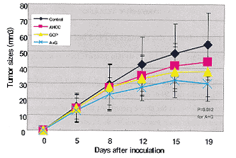
2) PC-3 bearing nude mice
The male, 5 weeks old nude mice were used in this study. 32 mice were divided into 4 groups and after housing for one week, all the mice were inoculated subcutaneously with PC-3 cells of 3 millions per mouse in 0.2 ml PBS mixed with 0.1 ml metrigel. From day 1, the four groups of control, AHCC, GCP and AHCC plus GCP were daily treated orally with water, 10% AHCC, 10% GCP and 10% AHCC mixed with 10% GCP at the volume of 0.1ml/10g body weight, respectively. The tumor sizes were measured every 3 or 4 days and calculated with the formula of length x width x height x 0.52.
As shown in Fig. 8, on the day 10 after treatment, the co-administration of AHCC and GCP inhibited the tumor growth significantly compared with the control group, even the tumor sizes were same among all four groups on day 5.
Fig. 8 Tumor size changes in PC-3 bearing nude mice
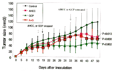
From the day 19, we stopped treating AHCC to AHCC group and treating GCP to AHCC plus GCP group, but kept treating GCP and AHCC to GCP and AHCC plus GCP groups, respectively. Doing so made us have the possibility to observe the real influences of AHCC and GCP to tumors in both prevention and treatment.
When the tumor sizes in AHCC and AHCC plus GCP groups became big near those in the control group after stopping AHCC and GCP to the two groups on the day 33.
Fig. 9 Tumor size changes in PC-3 bearing nude mice
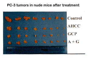
However, we treated these mice with AHCC and GCP again for another 10 days, the tumor sizes went down again (Fig.0). On the day 50 of the treatment, we sacrificed all the mice and took out the tumor tissues. The final tumors are shown in the above picture. At the end of the treatment, AHCC, GCP and AHCC plus GCP groups appeared to have much smaller tumors than control group (Fig. 10).
Fig. 10 PC-3 tumors in nude mice after treatment
3) The mechanisms of the effects of AHCC and GCP on tumors
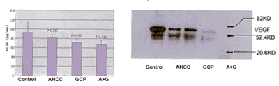
A. Down-regulating VEGF levels in tumor cells and tumor tissues
PC-3 cell line was cultured in RPMI1640 medium for 24 hrs, then, AHCC, GCP or AHCC plus GCP were added into the medium to the final concentration of 200 � g/ml medium. After culturing for another 48 hrs, the culture medium was collected and the proteins in the tumor cells were prepared for measuring VEGF by ELISA and Western Blotting, respectively. The results appeared as follows.
Fig. 11 VEGF expression in PEC tumor cell
(left: ELISA, right: Western Blotting)AHCC and GCP treatment down-regulated VEGF levels, but the co-administration of AHCC and GCP showed activity more obviously.
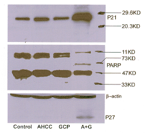
B. Up-regulating the protein expressions related to tumor inhibitory genes
In this study, p21, p27 and PARP proteins were extracted from tumor tissues and were checked with Western blotting.
Fig. 12 The protein expressions related to tumor inhibitory
As shown in the pictures above, the co-administration of AHCC and GCP regulated these genes that control the apoptosis happening in tumor tissues
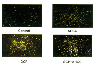
Fig. 13 Apoptosis in PC-3 tumor tissues (TUNEL)
Seen from above photographs, apoptosis happened only in approximately 1-3% of tumor cells in the control group,. but about 30% cells showed to be apoptosis cells in AHCC-treated group. In GCP and AHCC plus GCP-treated groups, near 50% tumor cells became apoptotic.
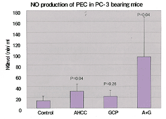
C. Enhancing the immune system
When the PC-3 bearing mice were sacrificed, PEC (peritoneal exudate cells) was collected and cultured in phenol red free RPMI1640 medium with 500ng/ml LPS for 36 hours. NO (nitrogen oxide) production was assayed by using Gries Reagent. As shown in Fig. 14, AHCC treatment increased NO production compared with the control group, but the co-administration showed a much stronger effect than the AHCC-treated group.
Fig. 14 NO production of PEC in PC-3 bearing mice
As a summary, AHCC and GCP appeared obviously inhibitory effects on prostate cancer cells, but the co-administration of AHCC and GCP showed synergistic effects to the cancer. The mechanisms might be:
- Down-regulate VEGF expression in serum, tumor cells and tissues.
- Up-regulate p21, p27 and PARP expression.
- Enhance immune system.
- Induce apoptosis in tumor cells and tumor tissues.
To explain why the co-administration of AHCC and GCP show synergistic effects on cancers, it can be thought that, at first, GCP up-regulates tumor-inhibitory genes and down-regulates VEGF levels so that apoptosis happens in tumors. After tumors become smaller, the tumor will be cleared by the immune system enhanced by AHCC.
Home | Product Report | News | Home
![]()
GCPresearch.com P.O. Box 311 Rye, NY 10580
Tel: (914) 251-0255 E-fax: (775) 599-7918
Email: [email protected]